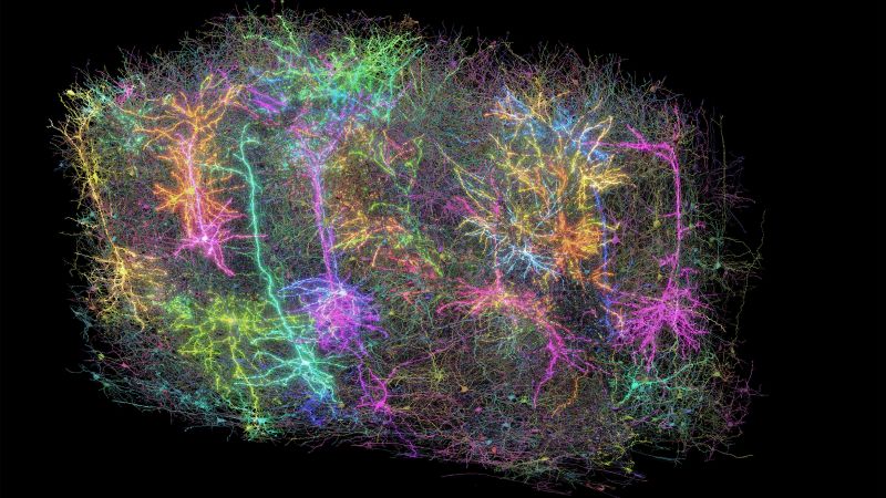
In a groundbreaking achievement within the realm of neuroscience, a team of dedicated researchers has successfully created the first highly detailed three-dimensional map of a mouse brain, capturing the intricate makeup of 84,000 neurons and more than 500 million synapses. The project, which is the outcome of nearly a decade of collaborative research involving 150 scientists from 22 different institutions, involved meticulous imaging and analysis, culminating in a pioneering resource that could enhance our understanding of human neurological disorders such as Alzheimer’s and Parkinson’s disease.
The map is constructed from a minuscule slice of brain tissue roughly the size of a grain of sand, yet this tiny portion contains an expansive network of 3.4 miles (approximately 5.4 kilometers) of neuronal wiring. This endeavor was spearheaded by the Allen Institute for Brain Science in collaboration with the Baylor College of Medicine and Princeton University. Remarkably, the depth of data generated from the project amounts to 1.6 petabytes, which, to visualize, is equivalent to an astonishing 22 years of continuous high-definition video.
The intricate details of the mapping process began with researchers at Baylor College of Medicine using specialized microscopes to monitor brain activity within a one-cubic-millimeter segment of a laboratory mouse’s visual cortex. This area is crucial as it processes visual information. The team ensured the mouse was awake, engaging it with stimuli—showing the animal clips from films like “The Matrix” and “Mad Max: Fury Road,” as well as adrenaline-inducing YouTube videos of extreme sports. This careful observation allowed researchers to capture baseline activity during specific visual processing tasks.
After euthanizing the mouse, the Allen Institute’s research team meticulously sliced the brain into over 28,000 ultra-thin layers, each measuring one-400th the width of a human hair. A transformative machine facilitated this slicing process, which required precision and constant oversight to ensure data integrity. Princeton researchers then employed machine learning algorithms to aid in the mapping process, tracing each neuron’s pathway through the network and segmenting them for detailed analysis.
Dr. Forrest Collman, the associate director of data and technology at the Allen Institute, expressed a sense of wonder at the beauty unveiled through the project. He likened the visual intricacies of neurons to the mesmerizing images of distant galaxies, emphasizing the awe-inspiring nature of discovering the brain’s architecture.
The implications of this research reach far beyond mere understanding; they offer potential avenues for diagnosing and treating various brain-related conditions. The mouse brain connectome—mapping how neurons connect—is viewed as a vital first step toward unraveling the complexities of human cognition and the neural disruptions that can lead to disorders. The idea is akin to having a circuit diagram for a malfunctioning radio, which significantly aids in pinpointing the flaws for correction.
Moving forward, the ambition of mapping the entire mouse brain connectome presents a significant challenge. Current estimates suggest the feasibility of achieving this in three to four years, but researchers also acknowledge the formidable hurdles in mapping the human connectome at the same resolution. The human brain is approximately 1,500 times larger than a mouse brain, which brings forth a host of technical and ethical challenges.
A noteworthy aspect of the research dives into the neocortex—a region generally associated with high cognitive functions. Both Dr. Mariela Petkova and Dr. Gregor Schuhknecht from Harvard, who did not participate in the study directly, highlighted that understanding this area plays a key role in grasping how sensory perception and decision-making are achieved by the mammalian brain. Ultimately, as advancements continue in this elucidating work, the mapping of the mouse brain stands not only as a scientific feat but also as a beacon of hope for future therapies targeting complex brain disorders.


